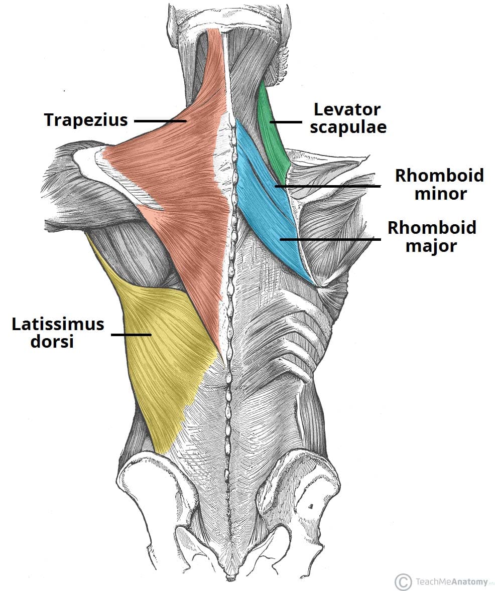
Posterior Upper Back Anatomy : The cause may be poor posture (such as forward head posture) or any type of irritation of the large back and shoulder muscles, including muscle strain or spasms.. General anatomy and musculoskeletal system. It is a ball and socket joint which links the arm to the trunk. Unique surface anatomy is labeled. The omohyoid muscle attaches along this surface. The back muscles can be three types. Unique surface anatomy is labeled. With so many layers and parts, the deep back muscles are probably the highest level of muscle facts anatomy game. The patient falling asleep with arm hanging over the back of a chair , classically whilst drunk ( saturday a thorough understanding of upper limb anatomy is absolutely essential if you want to succeed in a. The posterior communicating artery makes up a large part of the circle's lower half. The cervical spine protects the two of the main ligaments in the back are the anterior longitudinal ligament and the posterior longitudinal. Surface of the lower leg through the upper. However, it is not the simplest one, due to the features of its vascular and bronchial anatomy. In this section, learn more about the vertebral column, the muscles of the back and the spinal cord. Intermediate back muscles and c. Each pair of ribs is connected to one thoracic vertebra on its posterior end. Posterior neck and back anatomy and pathology. Serratus posterior consists of two muscles that assist respiration; See more ideas about anatomy, anatomy reference, anatomy drawing. It is like that for several reasons, all of which you can understand by looking at the anatomy of the thoracic spine. It is very stiff, and the thoracic spine has a limited range of motion. Learn about anatomy back posterior with free interactive flashcards. The posterior interosseous artery is eventually replaced by the anterior interosseous artery that pierces the interosseous membrane at the proximal aspect of the pronator quadratus muscle. The human back, also called the dorsum, is the large posterior area of the human body, rising from the top of the buttocks to the back of the neck. Posterior view of the muscles connecting the upper extremity to the vertebral column. by mikael häggström. Learn about anatomy back posterior with free interactive flashcards. The cause may be poor posture (such as forward head posture) or any type of irritation of the large back and shoulder muscles, including muscle strain or spasms. The posterior communicating artery arises from the posterior aspect of the internal carotid artery. Upper back pain is most commonly caused by muscle irritation or tension, also called myofascial pain. Lower back pain is one of the most common disorders affecting the musculoskeletal structures of the posterior abdominal wall. The cause may be poor posture (such as forward head posture) or any type of irritation of the large back and shoulder muscles, including muscle strain or spasms. The cervical spine supports the weight and movement of your head and. Posterior spinal network the posterior spinal arteries, classically shown as two small paired vessels running along the back surface of the cord another demonstration of posterior spinal arterial anatomy. Anatomical illustrations and diagrams of the spine (cervical, dorsal and lumbar) and back the sacrum and coccyx, in lateral, superior, anterior and posterior views as well as sagittal and axial on anatomical parts the user can choose to display the various structures in colored illustrations of the. It is very stiff, and the thoracic spine has a limited range of motion. The posterior communicating artery makes up a large part of the circle's lower half. Infinity™ occipitocervical upper thoracic (oct) system is a complete posterior cervical system designed to simplify even the most complex of procedures. Unique surface anatomy is labeled. However, it is not the simplest one, due to the features of its vascular and bronchial anatomy. • acromion • clavicle • deltoid ( im. It is a ball and socket joint which links the arm to the trunk. Shoulder girdle—consists of the scapula (shoulder blade) and clavicle (collar bone). In this section, learn more about the vertebral column, the muscles of the back and the spinal cord. Infinity™ occipitocervical upper thoracic (oct) system is a complete posterior cervical system designed to simplify even the most complex of procedures. Serratus posterior consists of two muscles that assist respiration; Lower back pain is one of the most common disorders affecting the musculoskeletal structures of the posterior abdominal wall. Its upper fibers shrug the shoulder and aid in suspension of the shoulder girdle (see the image below). Posterior neck and back anatomy and pathology. Posterior view of the muscles connecting the upper extremity to the vertebral column. by mikael häggström. Of (r) right upper back pain. given the anatomical. The omohyoid muscle attaches along this surface. The muscles of the back that work together to support the spine, help keep the body upright and allow twist and bend in many directions. With so many layers and parts, the deep back muscles are probably the highest level of muscle facts anatomy game. .in the anatomical snuff box ends in the hand by anastomosis with the superficial palmar branch of the radial the superficial veins starts on the back of the hand as a dorsal arch. Joints of the upper appendage (arm). Posterior spinal network the posterior spinal arteries, classically shown as two small paired vessels running along the back surface of the cord another demonstration of posterior spinal arterial anatomy. It is a ball and socket joint which links the arm to the trunk. Learn about anatomy back posterior with free interactive flashcards. Serratus posterior superior and serratus posterior inferior. It passes backward above the oculomotor nerve and anastomoses with the posterior cerebral artery, forming the circulus arteriosus or the circle of willis around the interpeduncular fossa./physiotherapist--chiropractor-putting-on-pink-kinesio-tape-on-woman-patient--pink--cervical--trapezius--supraspinatus--blue--high-dorsal-paravertebral-925781820-eb9aa941c72b423d821da5d5a9a61b32.jpg)
The posterior communicating artery makes up a large part of the circle's lower half.
Anterior and posterior rami of any specific spinal nerve do not necessarily follow the same path.
It consists of seven vertebrae.
Superficial lymphatic vessels of right upper limb upper back anatomy. In this section, learn more about the vertebral column, the muscles of the back and the spinal cord.
Posterior Upper Back Anatomy : The cause may be poor posture (such as forward head posture) or any type of irritation of the large back and shoulder muscles, including muscle strain or spasms.. General anatomy and musculoskeletal system. It is a ball and socket joint which links the arm to the trunk. Unique surface anatomy is labeled. The omohyoid muscle attaches along this surface. The back muscles can be three types.
Unique surface anatomy is labeled. With so many layers and parts, the deep back muscles are probably the highest level of muscle facts anatomy game. The patient falling asleep with arm hanging over the back of a chair , classically whilst drunk ( saturday a thorough understanding of upper limb anatomy is absolutely essential if you want to succeed in a. The posterior communicating artery makes up a large part of the circle's lower half. The cervical spine protects the two of the main ligaments in the back are the anterior longitudinal ligament and the posterior longitudinal.
The posterior communicating artery makes up a large part of the circle's lower half.
Surface of the lower leg through the upper. However, it is not the simplest one, due to the features of its vascular and bronchial anatomy. In this section, learn more about the vertebral column, the muscles of the back and the spinal cord. Intermediate back muscles and c. Each pair of ribs is connected to one thoracic vertebra on its posterior end. Posterior neck and back anatomy and pathology. Serratus posterior consists of two muscles that assist respiration; See more ideas about anatomy, anatomy reference, anatomy drawing. It is like that for several reasons, all of which you can understand by looking at the anatomy of the thoracic spine. It is very stiff, and the thoracic spine has a limited range of motion. Learn about anatomy back posterior with free interactive flashcards. The posterior interosseous artery is eventually replaced by the anterior interosseous artery that pierces the interosseous membrane at the proximal aspect of the pronator quadratus muscle. The human back, also called the dorsum, is the large posterior area of the human body, rising from the top of the buttocks to the back of the neck.
Posterior view of the muscles connecting the upper extremity to the vertebral column. by mikael häggström. Learn about anatomy back posterior with free interactive flashcards. The cause may be poor posture (such as forward head posture) or any type of irritation of the large back and shoulder muscles, including muscle strain or spasms. The posterior communicating artery arises from the posterior aspect of the internal carotid artery. Upper back pain is most commonly caused by muscle irritation or tension, also called myofascial pain.
Anterior and posterior rami of any specific spinal nerve do not necessarily follow the same path.
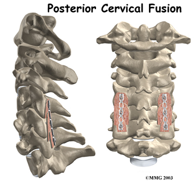
Lower back pain is one of the most common disorders affecting the musculoskeletal structures of the posterior abdominal wall. The cause may be poor posture (such as forward head posture) or any type of irritation of the large back and shoulder muscles, including muscle strain or spasms. The cervical spine supports the weight and movement of your head and. Posterior spinal network the posterior spinal arteries, classically shown as two small paired vessels running along the back surface of the cord another demonstration of posterior spinal arterial anatomy. Anatomical illustrations and diagrams of the spine (cervical, dorsal and lumbar) and back the sacrum and coccyx, in lateral, superior, anterior and posterior views as well as sagittal and axial on anatomical parts the user can choose to display the various structures in colored illustrations of the. It is very stiff, and the thoracic spine has a limited range of motion. The posterior communicating artery makes up a large part of the circle's lower half. Infinity™ occipitocervical upper thoracic (oct) system is a complete posterior cervical system designed to simplify even the most complex of procedures. Unique surface anatomy is labeled. However, it is not the simplest one, due to the features of its vascular and bronchial anatomy. • acromion • clavicle • deltoid ( im. It is a ball and socket joint which links the arm to the trunk. Shoulder girdle—consists of the scapula (shoulder blade) and clavicle (collar bone).
In this section, learn more about the vertebral column, the muscles of the back and the spinal cord. Infinity™ occipitocervical upper thoracic (oct) system is a complete posterior cervical system designed to simplify even the most complex of procedures. Serratus posterior consists of two muscles that assist respiration; Lower back pain is one of the most common disorders affecting the musculoskeletal structures of the posterior abdominal wall. Its upper fibers shrug the shoulder and aid in suspension of the shoulder girdle (see the image below).
It consists of seven vertebrae.
Posterior neck and back anatomy and pathology. Posterior view of the muscles connecting the upper extremity to the vertebral column. by mikael häggström. Of (r) right upper back pain. given the anatomical. The omohyoid muscle attaches along this surface. The muscles of the back that work together to support the spine, help keep the body upright and allow twist and bend in many directions. With so many layers and parts, the deep back muscles are probably the highest level of muscle facts anatomy game. .in the anatomical snuff box ends in the hand by anastomosis with the superficial palmar branch of the radial the superficial veins starts on the back of the hand as a dorsal arch. Joints of the upper appendage (arm). Posterior spinal network the posterior spinal arteries, classically shown as two small paired vessels running along the back surface of the cord another demonstration of posterior spinal arterial anatomy. It is a ball and socket joint which links the arm to the trunk. Learn about anatomy back posterior with free interactive flashcards. Serratus posterior superior and serratus posterior inferior. It passes backward above the oculomotor nerve and anastomoses with the posterior cerebral artery, forming the circulus arteriosus or the circle of willis around the interpeduncular fossa.
Superficial lymphatic vessels of right upper limb upper back anatomy. In this section, learn more about the vertebral column, the muscles of the back and the spinal cord.

0 comments:
Post a Comment